

The approach, at a glance.
These consultation points summarize procedure-specific descriptions from the official Korean detail page. Suitability, technique, risks and recovery require individual clinical assessment.
Adhesion-based method
The source describes encouraging adhesion between eyelid tissue and the eye-opening muscle.
Small openings
Small openings and suture knots are described in the source.
Eyelid assessment
The source discusses thin eyelids and the need to assess drooping or tissue removal before selecting a method.
Choosing a method
The FAQ explains that the preferred fold alone does not determine the method; fat, skin and ptosis also matter.
Process described in the source
Consultation
The source begins with an assessment of the eyes.
Fold design
The source describes planning the fold starting point and position.
Adhesion approach
The source describes a procedure intended to encourage adhesion.
Suture knots
The source then discusses knots used to support the fold.
This diagram follows the source’s explanation order. It is not an individual operative plan or a promised result.
How the approach works.
The surgeon plans the crease and uses internal fixation to encourage adhesion between the tissues. It is different from removing excess skin through a longer incision. A crease can loosen, and the method is not suitable for every eyelid.
Upper-eyelid movement assessment

A surface view shows the upper eyelid crease and lid margins around an intact eye.
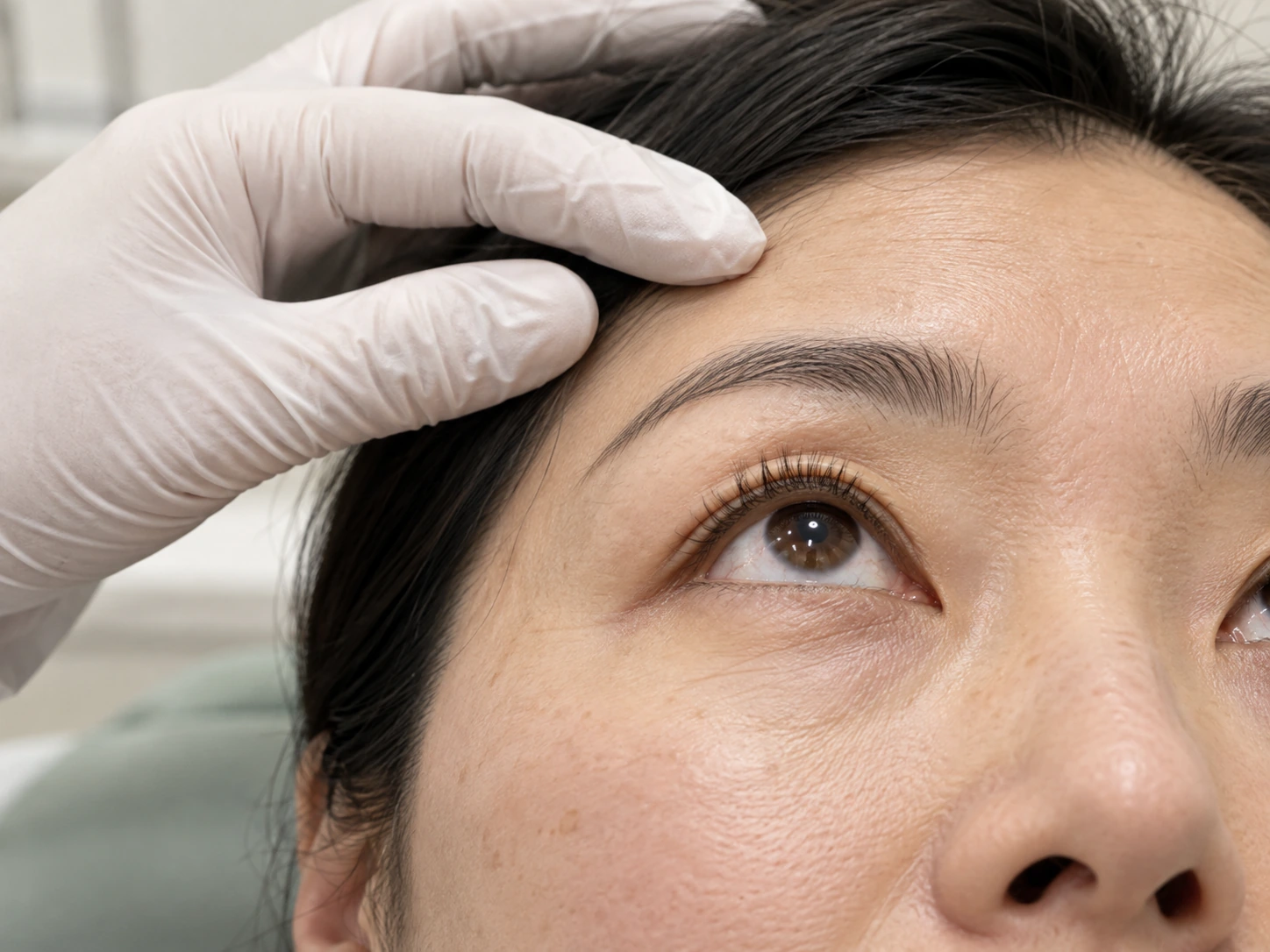
Upper-eyelid position and movement are assessed while brow activity is considered separately. The levator and its aponeurosis are explained in the accompanying text.
The levator muscle runs above the eyeball and continues toward the upper eyelid through its aponeurosis.
Upper Eyelid Planning Marks
Illustrative marks along an intact upper eyelid crease during planning. They are not a prescribed placement guide and do not show internal fixation or a surgical result.
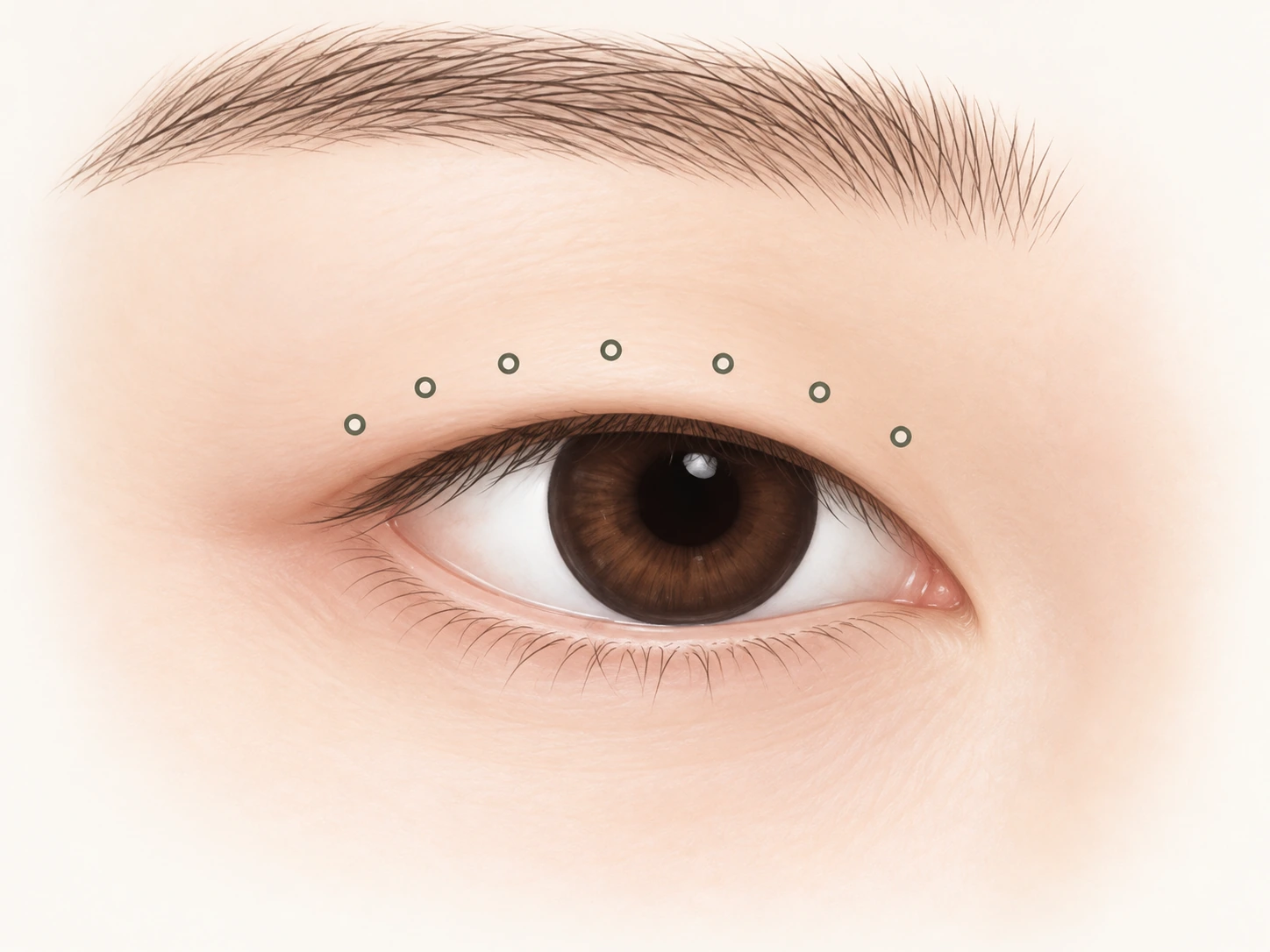
Planning the crease
The crease position is planned after assessing the eyelid.
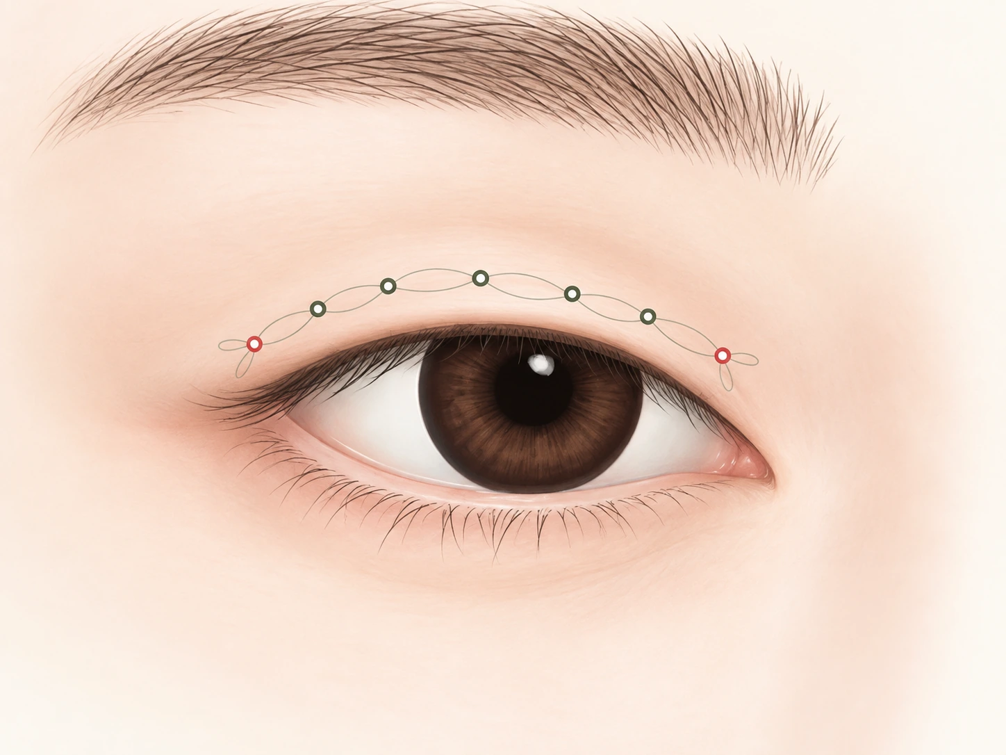
Internal fixation
Internal fixation is used to encourage adhesion between tissues.
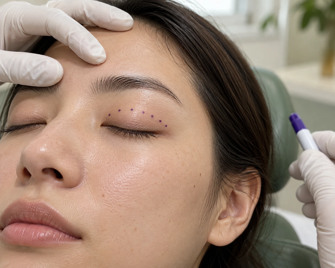
Illustrative marks along an intact upper eyelid crease during planning. They are not a prescribed placement guide and do not show internal fixation or a surgical result.
Treatment illustration; not a patient result.
What the consultation considers.
Eyelid thickness
Excess skin
Previous crease surgery
This is a starting point for a consultation, not an individual treatment recommendation.
Structures to discuss
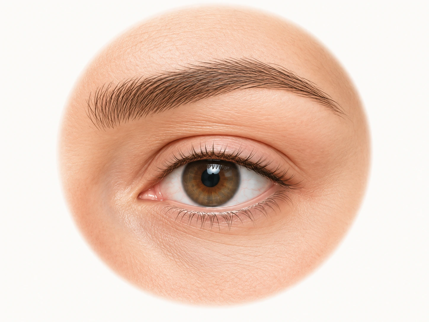
The eyebrow lies above the upper eyelid crease; their positions vary between individuals.
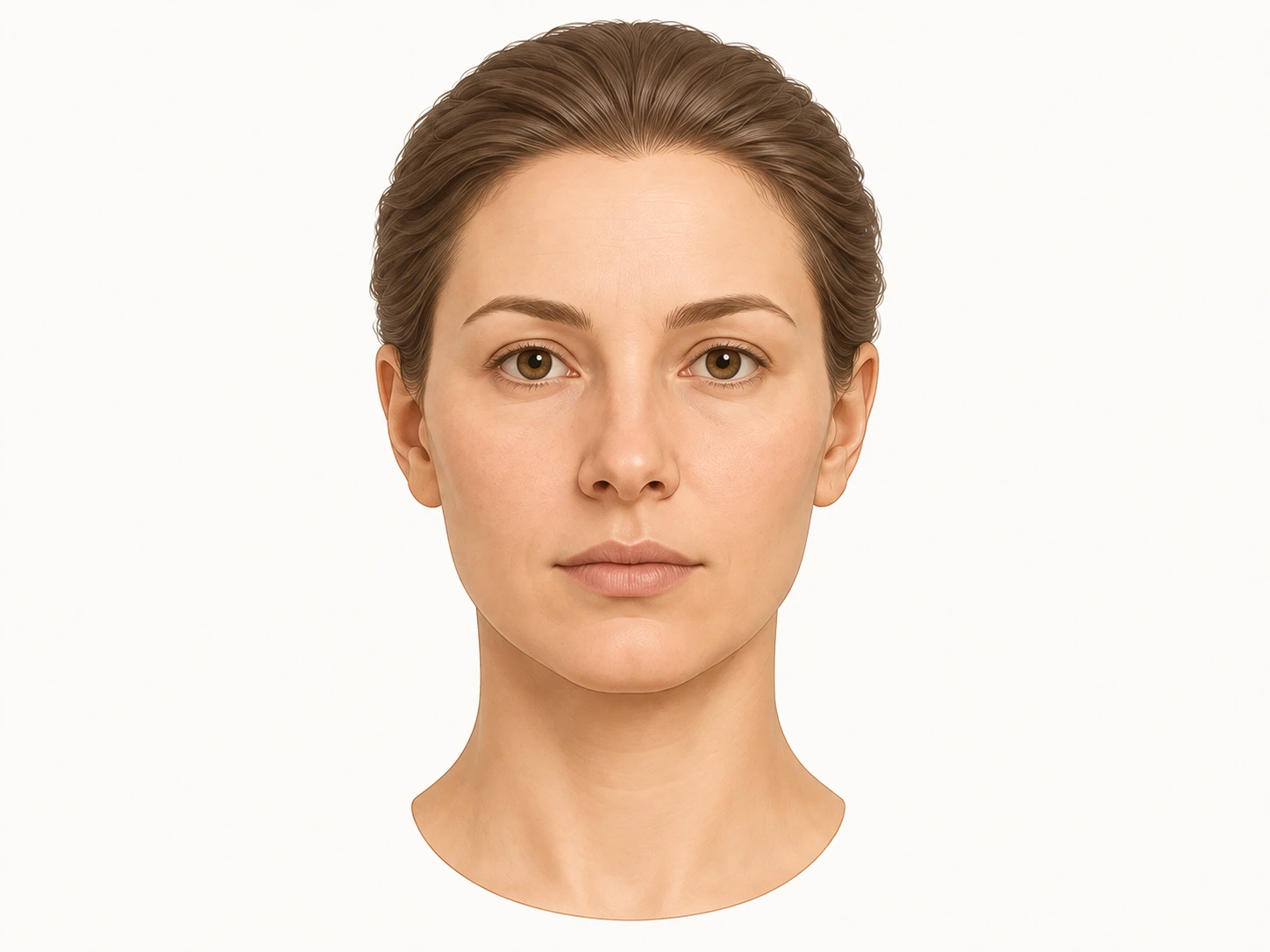
A neutral view of the same fictional teaching model, showing the eyes, nose, lips and chin.
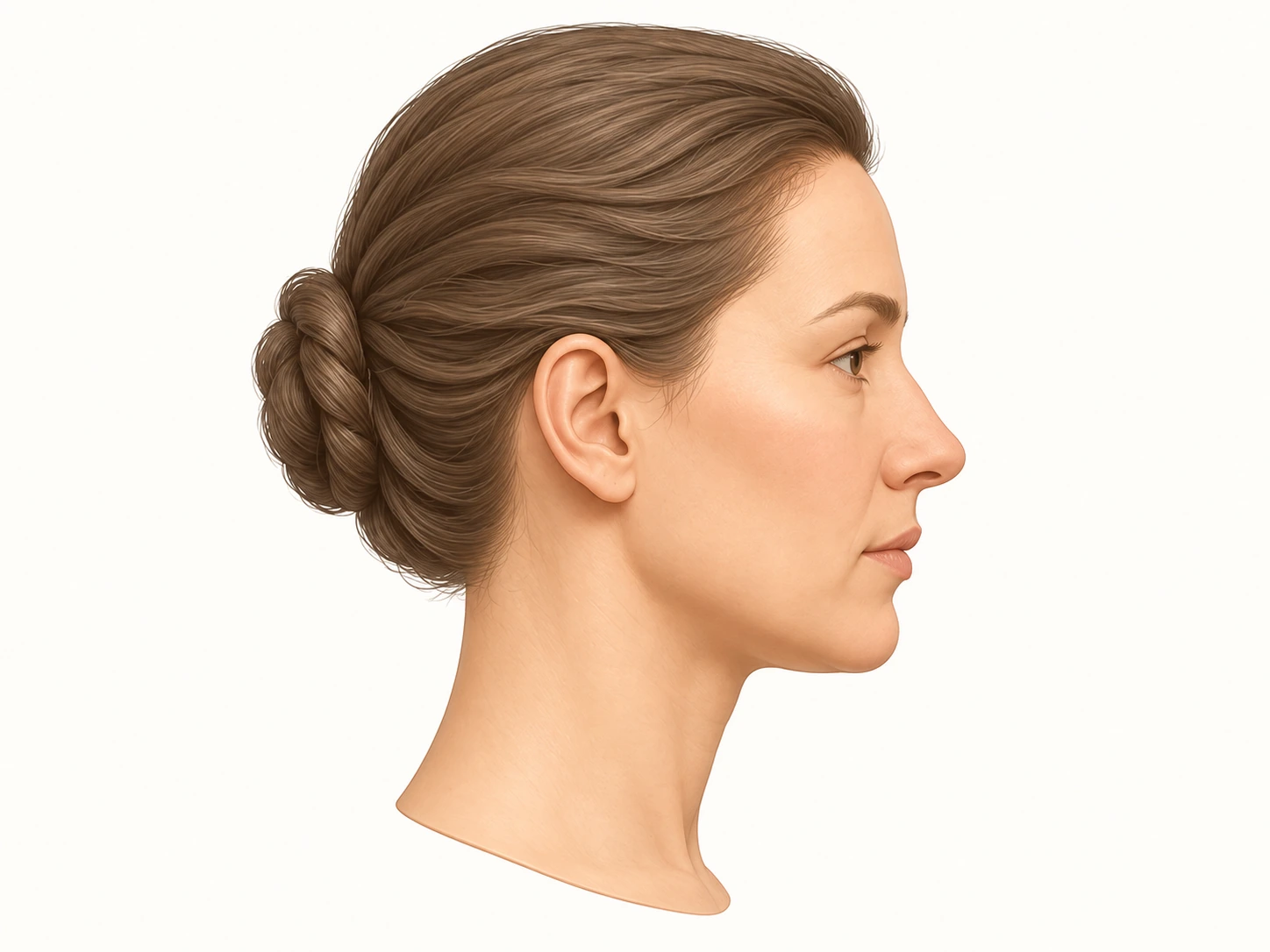
The side view shows the forehead, nose, lips, chin and jaw outline without changing the model.
Recovery, risks & limits.
Some reactions can be temporary; complications can also require treatment. This is not an exhaustive list.
Swelling, bruising, discomfort and scarring can occur.
Dryness, difficulty closing the eyes, asymmetry or a need for revision should be discussed.
Bleeding, infection and rare serious changes in vision require specific discussion with the surgeon.
Your timeline is individual.
Procedure time, anaesthesia, follow-up and return travel depend on your examination and the final plan. Confirm them before paying for flights or accommodation.
Recovery and results vary. Your clinician should explain the procedure-specific risks and aftercare before you decide.
If you develop severe or worsening symptoms after treatment, seek medical attention rather than waiting for an online reply.
Your questions, answered.
Is a non-incisional approach always possible?
No. The official clinic information distinguishes thin eyelids from cases that need skin or fat removal or correction of weak eye opening. The examination determines the approach.
How long should I stay in Korea?
Procedure time, anaesthesia, follow-up and return travel depend on your examination and the final plan. Confirm them before paying for flights or accommodation.
Can I choose the procedure before an examination?
You can explore your options first. The final plan requires an examination, a discussion of alternatives and informed consent. Choosing no treatment is also an option.
How is the cost decided?
The clinic confirms the proposed method, treatment area and product or equipment before giving an individual quote. Ask what is included in follow-up care.
What should I disclose before treatment?
Tell the clinician about previous procedures, implants, medication, allergies and health conditions. Bring available details of earlier injections or surgery.
Information sources
Information checked against official websites on 30 October 2026
Official Korean sourceOfficial English sourceOfficial Traditional Chinese sourceBefore & After
Move the divider to compare the original photographs.
Natural-adhesion double eyelid + ptosis correction + inner/upper canthoplasty
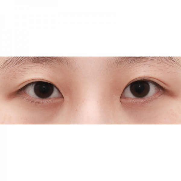
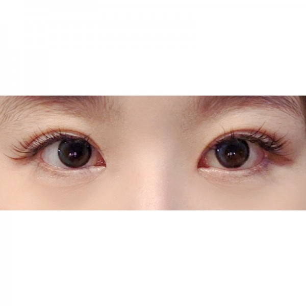 BeforeAfter
This case includes multiple procedures; the photographs do not isolate the effect of this treatment.
View photo source ↗Original photographs published on the official Korean website. Individual results vary. The source does not state when the follow-up photograph was taken.
Official case gallery ↗Some photographs on the official website require sign-in.